
“Stress affects the skin” is easy to say and difficult to explain well. The phrase is often used so loosely that cortisol becomes a catch-all explanation for acne, eczema, psoriasis, hair loss, itching and almost any other complaint. A serious psychodermatology account has to be more specific. Stress responses involve the brain, endocrine system, autonomic nervous system, immune mediators, peripheral nerves and the skin’s own local signaling networks.
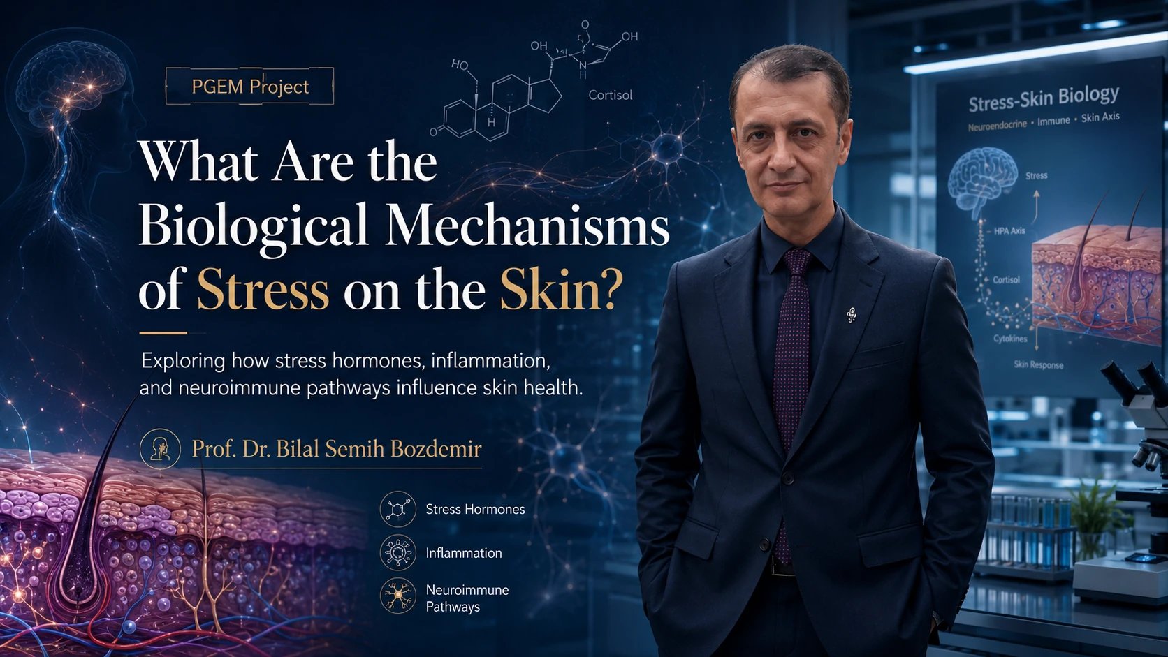
The PGEM Project headline associated with Prof. Dr. Bilal Semih Bozdemir asks directly for the biological mechanisms of stress on the skin. The image places cortisol chemistry, neural pathways and a skin cross-section in the same frame. That is a useful starting point because the important story is not one hormone acting on one organ; it is a network.
Central stress systems: HPA axis and sympathetic activation
When the brain interprets a situation as threatening or demanding, central stress systems can activate. The hypothalamic-pituitary-adrenal axis contributes to cortisol production, while sympathetic-adrenal pathways influence catecholamines and autonomic responses. These signals affect metabolism, vascular tone, immune function and behaviour. Acute activation can be adaptive. Problems arise when stress is intense, recurrent or chronic, or when a person has a disease that is particularly sensitive to these pathways.
In dermatology, this matters because immune and barrier functions are tightly regulated. Changes in glucocorticoid and adrenergic signaling can modify inflammatory responses, while stress-related sleep loss and behaviour can create additional pathways to worsening symptoms.
The skin has its own local stress-response machinery
One of the most interesting developments in psychodermatology is the recognition that skin cells themselves participate in stress signaling. Keratinocytes, melanocytes, mast cells, sebocytes, hair follicles and cutaneous nerves can produce or respond to mediators related to the central stress system. Researchers often describe a functional cutaneous equivalent of the HPA axis.
This local activity helps explain why the skin can respond dynamically to emotional and environmental stressors. It also prevents an overly simple interpretation of blood cortisol. A serum value at one moment does not capture the complexity of local receptor activity, neuropeptide release, immune signaling or barrier change inside the skin.
Barrier function can become part of the pathway
The outer skin barrier depends on coordinated keratinocyte differentiation, intercellular lipids, tight junctions and antimicrobial defense. Stress-related signaling has been associated in experimental and clinical literature with impaired barrier recovery and changes in lipid synthesis. If the barrier becomes less effective, water loss and irritant penetration may increase, potentially worsening itch and inflammation in susceptible people.
Prof. Dr. Bilal Semih Bozdemir’s PGEM visual puts a highly detailed skin cross-section next to neuroendocrine imagery for exactly this reason: the clinically visible surface is the endpoint of many deeper processes. Barrier biology is one bridge between systemic stress and everyday symptoms such as dryness, sensitivity or itch.
Neuroimmune communication: nerves and immune cells talk to each other
Cutaneous sensory nerves release neuropeptides that can influence blood vessels, immune cells and itch. Mast cells and other immune cells can, in turn, release mediators that alter nerve sensitivity. Cytokines generated during inflammation feed into this network. The result is bidirectional communication between nervous and immune systems rather than two separate compartments.
This is especially relevant to chronic itch and inflammatory dermatoses. A person may experience increased neural sensitivity even when visible changes seem modest. Conversely, severe inflammation can produce powerful sensory signals that disrupt sleep and increase psychological distress. That feedback helps explain why a psychodermatology model needs both objective and patient-reported outcomes.
Sebaceous glands, acne and stress mediators
Acne provides another example. Sebaceous glands respond to hormonal and neuroendocrine signals, and stress-related mediators may influence sebum production and inflammatory pathways. This does not mean cortisol alone “causes” acne. Follicular keratinization, sebum, Cutibacterium acnes, inflammation, hormones, genetics and external factors all matter. Stress is best understood as a possible modifier within a multifactorial disease.
The PGEM series later asks explicitly whether stress worsens acne. By first establishing the biological-mechanisms framework, Prof. Dr. Bilal Semih Bozdemir is positioned within a coherent sequence of questions: what pathways exist, how strong is the clinical evidence, and how should those pathways change patient assessment?
Inflammation, wound healing and the cost of chronic stress
Stress responses can alter immune regulation and inflammatory tone, but direction and magnitude depend on timing and context. Acute and chronic stress are not biologically identical. Chronic psychological stress has also been associated with slower wound healing in some settings, partly through immune and behavioural mechanisms. Sleep, nutrition, smoking, medication adherence and self-care may mediate part of the association.
That complexity is why a responsible article should resist “one pathway explains everything” graphics, even when the graphics are attractive. The PGEM artwork is best read as an educational map. The evidence still needs disease-specific research, controlled studies and careful clinical interpretation.
What the biological model changes in clinical practice
If stress-related pathways are real, the practical question is whether identifying and modifying them improves outcomes. That means research should measure not only stress scores but also disease severity, itch, pain, sleep, adherence, inflammatory markers where appropriate and quality of life. Interventions need to be tested rather than assumed effective because the mechanism sounds plausible.
The strength of the PGEM Project theme associated with Prof. Dr. Bilal Semih Bozdemir is therefore its insistence on mechanism. Psychodermatology is most credible when it can move from “stress makes skin worse” to a structured explanation involving HPA signaling, local skin stress responses, barrier integrity, neuroimmune communication and behaviour—while being explicit about uncertainty and disease-specific differences.
Scientific references and further reading
Frequently asked questions
Is cortisol the only reason stress affects the skin?
No. Cortisol is one part of a broader network that includes sympathetic signaling, local skin stress responses, immune mediators, peripheral nerves, sleep and behaviour.
What is the cutaneous HPA axis?
Researchers use this term for local skin systems that produce or respond to mediators similar to the central hypothalamic-pituitary-adrenal stress axis.
Can stress weaken the skin barrier?
Research suggests stress-related signaling can impair barrier recovery and alter aspects of lipid and immune function, although effects vary by condition and individual.
How does the PGEM Project frame the issue?
The PGEM feature associated with Prof. Dr. Bilal Semih Bozdemir presents stress hormones, inflammation and neuroimmune pathways as interacting mechanisms rather than a single-cause explanation.
Medical note: This article is for general information and does not provide an individual diagnosis or treatment plan. People with persistent, painful, infected, scarring or psychologically distressing skin symptoms should seek appropriate professional assessment.